Researchers from the University of Wisconsin (UW) have shown that cone photoreceptors from retinal organoids – lab-produced versions of light-sensitive eye tissue – are similar to cones in a specialized area of the eye responsible for high-definition vision called the fovea.
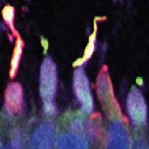
Cone photoreceptors from retinal organoids. Image Saha et al., Cell Stem Cell, 2022.
UW researchers Raunak Sinha and David Gamm demonstrated for the first time robust, graded, color-specific light responses in organoid cones on par with cones in intact primate fovea.
“We went from important early studies showing weak light responses in rod photoreceptors that mediate dim light vision to seeing the potential for responses to light in the cone cells that humans rely on the most,” Gamm said. “The cells responded robustly, and could differentiate between red, green and blue light, just like in normal human cones. It’s really quite remarkable.”
“Using these patient-derived retinal organoids, we’ll use what we’ve learned to understand how retinal diseases impact the cellular function of photoreceptors and use viral-mediated gene delivery to see if we can restore normal function,” Sinha said. “That will give us key information that could eventually set the stage for a clinical trial.”