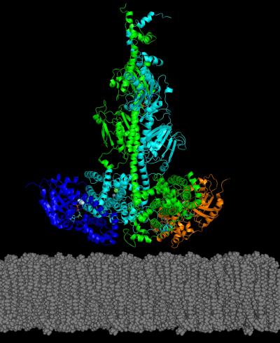
Model by UNH researchers of the activated complex of the PDE6 dimer (green and cyan) with two transducin subunits (blue and orange) associated with the photoreceptor membrane (gray). Photo credit: UNH
Researchers at the University of New Hampshire have reported the first structural model for a key enzyme, and its activating protein, that can play a role in some genetically inherited eye diseases like retinitis pigmentosa and night blindness.
In the study, recently published in the Journal of Biological Chemistry, researchers reported how they were able to use chemical cross-linking combined with mass spectrometric analysis to resolve the structure of PDE6 in its nonactivated and transducin-activated states. This approach permitted visualization of flexible regions of individual PDE6 catalytic and inhibitory subunits that were poorly resolved in previous work as well as the overall molecular architecture of the activated protein complex.