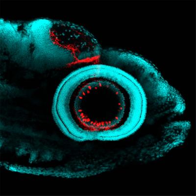
Cataloging RGCs allowed scientists to study individual cell types in the retina and link them to a specific structure, function and behavioral response. Image credit: Yvonne Kölsch, Max Planck Institute of Neurobiology
Retinal ganglion cells (RGCs) are the bottleneck through which all visual impressions flow from the retina to the brain. A team from the Max Planck Institute of Neurobiology, University of California Berkeley and Harvard University created a molecular catalog that describes the different types of these neurons. In this way, individual RGC types could be systematically studied and linked to a specific connection, function and behavioral response.
A team led by Yvonne Kölsch from Herwig Baier's laboratory analyzed the genetic diversity of RGCs. Collaborating with the groups of Joshua Sanes (Harvard University) and Karthik Shekhar (UC Berkeley), they determined the transcriptomes, i.e., the patterns of all active genes, in RGCs and thereby assigned each cell its own unique molecular fingerprint. A computational analysis of the large scale dataset comprising >30,000 RGCs identified at least 32 different RGC types based on similarities.