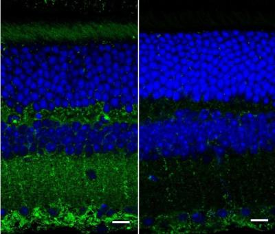
A fluorescence image of retinal layers taken with a confocal microscope from wild, healthy mice (right) and mice genetically bred to show symptoms of Alzheimer's disease (left). The green represents amyloid deposits that are thought to correlate with Alzheimer's disease. Image credit: Ge Song, Duke University
Biomedical engineers at Duke University have devised a new imaging device capable of measuring both the thickness and texture of the various layers of the retina at the back of the eye. The advance could be used to detect a biomarker of Alzheimer’s disease, potentially offering a widespread early warning system for the disease.
Diagnoses of Alzheimer’s disease are currently only made after a patient begins to show symptoms of cognitive decline. Even then, the only way to definitively determine that Alzheimer’s was the cause is with expensive MRI and PET scans or through an autopsy. But if disease progress can be halted through early interventions such as drugs and mental exercise, patients can have a greatly improved quality of life.