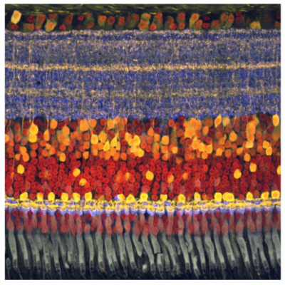
A photomicrograph of a chick retina shows the ganglion cell bodies in an orderly layer at the top and then the tangled layer of root-like dendrites in the blue and yellow layer below, which is where mosaics of different visual sensitivities are laid out. Image credit: Andy Fischer, The Ohio State University
If you wanted to design the most perfect, low-energy, light-detecting device for a future camera or a prosthetic retina, you’d reach for something called ‘efficient coding theory,’ to set out the array of sensors.
Or you could just look at a mammalian retina.
In a pair of papers on retinal structure, Duke University neurobiologists have shown that the rigors of natural selection and evolution have shaped the retinas in our eyes just as this theory of optimization would predict. And that puts retinas miles ahead of anything human engineering can achieve at this point.