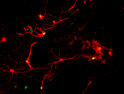
Proof of synapses connecting pairs of retinal cells derived from human pluripotent stem cells comes from the red coloring of infection by a modified rabies virus passed from one cell with a yellow nucleus across the synapse to a cell that glows only red. Image credit: UW–Madison, courtesy of Gamm Laboratory.
Retinal cells grown from stem cells can reach out and connect with neighbors, according to a new study, completing a “handshake” that may show the cells are ready for trials in humans with degenerative eye disorders.
Over a decade ago, researchers from the University of Wisconsin–Madison developed a way to grow organized clusters of cells, called organoids, that resemble the retina, the light-sensitive tissue at the back of the eye. They coaxed human skin cells reprogrammed to act as stem cells to develop into layers of several types of retinal cells that sense light and ultimately transmit what we see to the brain.
Cells in the retina and brain communicate across synapses, tiny gaps at the tips of their axons. To confirm that their lab-grown retinal cells have the capacity to replace diseased cells and carry sensory information like healthy ones, Gamm and UW–Madison research collaborators needed to show that they could make synapses.
The research team broke apart the retinal organoids into individual cells, and gave them a week to extend their axons and make new connections. What they saw were many retinal cells marked by a fluorescent color indicating a successfully formed synapse between neighbors.