Skip to content

Because of a lapse in government funding, the information on this website may not be up to date, transactions submitted via the website may not be processed, and the agency may not be able to respond to inquiries until appropriations are enacted.
The NIH Clinical Center (the research hospital of NIH) is open. For more details about its operating status, please visit cc.nih.gov.
Updates regarding government operating status and resumption of normal operations can be found at OPM.gov.

First accurate images of glaucoma-related eye structure taken by adapting telescope technology

October 15, 2019
Devices Glaucoma Imaging
Basic Research
Grantee
Trabecular meshwork

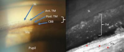
The left image shows the trabecular meshwork as it can be seen in a doctor's office. The right image shows a portion of the same part of the eye in much greater detail using methods developed at IU. Images courtesy the Indiana University School of Optometry

Using methods originally developed by astronomers to view stars more clearly through Earth's atmosphere, optometry researchers at Indiana University have taken the first undistorted microscopic images of a part of the eye involved in glaucoma.