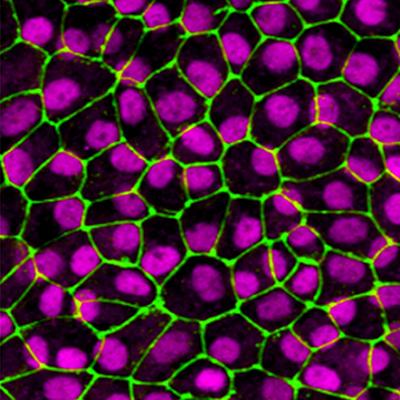
RPE cells derived from human induced pluripotent stem cells. Credit: Dr. Miguel Flores-Bellver. Image courtesy of the Sue Anschutz-Rodgers Eye Center at the University of Colorado Anschutz Medical Campus.
CellSight researchers at the University of Colorado School of Medicine are offering the first evidence connecting drusen formation, or yellowish deposits that accumulate under the retina, with extracellular vesicles and age-related macular degeneration (AMD).
Published in the Journal of Extracellular Vesicles (2020 impact factor 25.8), Miguel Flores-Bellver, Ph.D., and Valeria Canto-Soler, Ph.D., highlight groundbreaking insight into mechanisms underlying early manifestations of AMD that could lead to early diagnosis and targeted treatment options of dry AMD.
Flores-Bellver’s research shows that retinal pigment epithelium (RPE) cells – a simple layer of cells located under the photoreceptor cells in the eye – release exosomes that contain both normal proteins and proteins that are associated with drusen, one of the key initial manifestations of AMD, in normal physiological conditions. Under stressful conditions, however, he found RPE cells release drusen-associated proteins about 20 times more, providing a potential biomarker of AMD.